Objective: Engage in a discourse on the fluid mosaic model of membrane architecture.
Preface
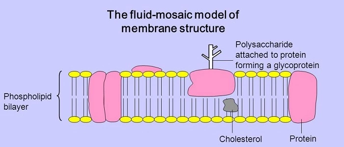
The fluid mosaic model describes a cell membrane characterized by a two-dimensional liquid with a heterogeneous composition. The cell membrane exhibits fluidity owing to its hydrophobic constituents, which contribute to its structure, allowing lipids and proteins inside the membrane to migrate laterally. The membrane has a more fluidic nature. The word mosaic, as used in the fluid mosaic model, denotes a composition created from diverse elements, so effectively validating its application in the current model. The cell membrane consists of several components, hence the term "mosaic" in the designation of the fluid mosaic model. The cell membrane exhibits fluidity owing to the existence of phospholipids that do not form bonds with one another. The phospholipid molecules include a head that is hydrophilic, orienting itself towards the outer surface of the cell membrane due to its attraction to water. The molecules migrate in two directions. The tail aids in dispersing the water that accumulates as a bilayer, rendering it non-polar and hydrophobic. The tails of phospholipids consist of fatty acids and exhibit continuous motion. The discovery of the bilayer occurred after the development of the fluid mosaic model. The procedure is conducted on an individual basis, making it less effective (Catala, 2012). The bilayer incorporates several proteins and chemicals, including cholesterol production. Proteins and several other substances traverse the plasma membrane owing to its viscosity, akin to that of vegetable oil at ambient temperature. The fluid mosaic model elucidates the structure of the plasma membrane. The cell membrane comprises several substances, each serving a specific function. Cholesterol embedded in the membrane bilayer provides stability, inhibiting solidification at lower body temperatures. The fluid mosaic model elucidates the plasma membrane of animal cells. The external layer of the cell membrane comprises carbohydrate chains that assemble into glycoproteins and glycolipids. The synthesis of carbohydrates varies across individuals, and their properties are contingent upon the individual's blood type (Leabu, 2013).

S.J. Singer and G.L. Nicolson developed the fluid mosaic model to elucidate the structure of the plasma membrane. The fundamental structure of the cell membrane was seen as crucial in elucidating the current data on the proteins and lipids inside the membrane, hence aiding future investigations (Nicolson, 2016). Despite its evolution throughout time, the model remains a relevant paradigm for elucidating the nano-structures of many intra- and cellular membranes seen in plant and animal cells. The fluid mosaic model describes the organization of particles inside the cell membrane. Meanwhile, new knowledge emerged on lipid rafts, proteins, and glycoproteins, which contributed to elucidating the membrane's structure. Recent insights into the cell membrane emerged from the principles underlying the Fluid Mosaic Model. The most recent information emphasizes the mosaic nature (Nicolson, 2016).
Cell organelles denote the many cellular components present in both plants and animals. A cell comprises many structures referred to as organelles. Cell organelles possess membranes that are integral to the cell, each exhibiting unique structures and functions (Morange, 2013). The cell membrane encapsulates all cellular components and protects the cell from external environments. It also assists in controlling the ions that enter and leave the cell. These cellular organelles operate in concert to ensure the proper functioning of the cell. The following cell organelles are mentioned below:
The endoplasmic reticulum is a network of membrane-bound channels filled with fluid. They are considered a transit mechanism facilitating the movement of materials inside the cell. The Endoplasmic Reticulum constitutes a component of the endomembrane system, extending from the nuclear envelope. The Endoplasmic Reticulum has two varieties: Rough Endoplasmic Reticulum and Smooth Endoplasmic Reticulum. The Rough Endoplasmic Reticulum consists of tubules, vesicles, and cisternae. They are distributed throughout the cell and facilitate protein synthesis. The Smooth Endoplasmic Reticulum serves as a storage facility. It facilitates the synthesis of steroids and lipids. It also aids in cellular detoxification. The Rough Endoplasmic Reticulum is characterized by many connected ribosomes, resulting in an irregular structure that validates its nomenclature. The Rough Endoplasmic Reticulum facilitates protein synthesis for export from the cell. The proteins are transported to the lumen inside the Endoplasmic Reticulum, where they undergo modification into a complex conformation. The lumen conveys the protein to the Smooth Endoplasmic Reticulum for further processing. The Smooth Endoplasmic Reticulum lacks ribosomes, contributing to its smooth texture. It is incapable of synthesizing proteins owing to the absence of ribosomes. The primary function of the Smooth Endoplasmic Reticulum is the synthesis of lipids and enzymes.

Source: (Dvorzhinskiy, 2018)
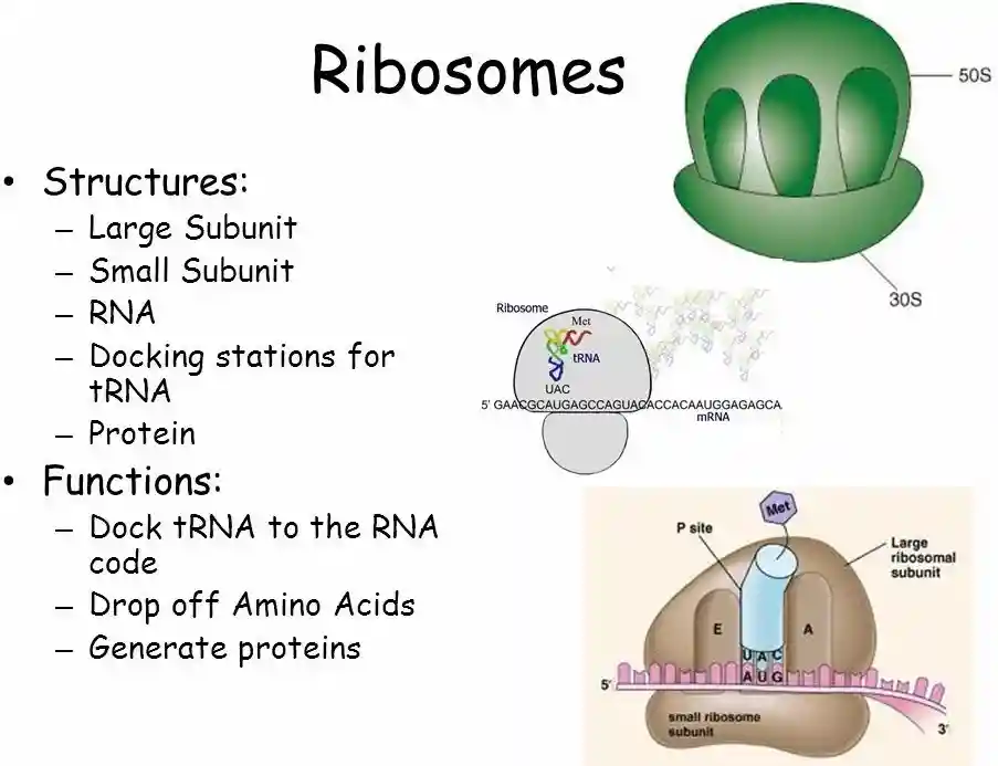
Ribosomes: It is a location where protein synthesis occurs, facilitating the production of proteins essential for the survival of live cells. It comprises many compounds. Ribosomes are present in all cell types, including bacterial and eukaryotic cells. Every cell need ribosomes for protein synthesis. Ribosomes have a small and a large component, composed of ribosomal RNA and ribosomal proteins. During protein synthesis, messenger RNA (mRNA) traverses the ribosome while amino acids bound to transfer RNA (tRNA) are delivered to the ribosome. Amino acids facilitate protein synthesis. The ribosomes are affixed to the Rough Endoplasmic Reticulum. They exist freely and suspend inside the cytoplasm. They are diminutive in nature and lack membranes. It comprises two-thirds ribonucleic acid and one-third protein. They are referred to either 70S, present in prokaryotes, or 80S, found in eukaryotes. S denotes density and dimensions. Ribosomes are present in the face, skin, hair, and eyes.

Source: ( Walker, n.d)
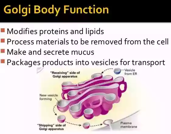
The Golgi apparatus, also known as the Golgi complex, is located in the cytoplasm of both plant and animal cells. It is a planar and stratified sac-like organelle that alters proteins and facilitates their packing. It is a collection of membranes that collaborates with the endoplasmic reticulum to alter proteins and carbohydrates. It typically comprises 3 to 20 cisternae, with an average of 6 cisternae. The Golgi apparatus is present in eukaryotic cells. It is encompassed by a membrane that varies in size across different locations. It is engaged in the manufacture, storage, and transportation of items derived from the Endoplasmic Reticulum. The membranes of the Golgi apparatus are folded, resulting in a pancake-like form. It serves as a repository for many vesicles generated by the Smooth Endoplasmic Reticulum. The Golgi apparatus facilitates the processing of proteins synthesized by the Endoplasmic Reticulum prior to their distribution inside the cell. The Endoplasmic Reticulum facilitates the entry of proteins into the Golgi apparatus from one side and their subsequent departure towards the plasma membrane of the cell from the other side. The fluid mosaic model is an established representation of the plasma membrane. The quantity of Golgi bodies inside a cell may vary based on its functions.

Source: ( Mandira and Kate, 2017)
Lysosomes: Lysosomes are often described as enzyme-filled sacs. It facilitates the digestion of various lipids and nucleic acids found inside a cell. The environment inside lysosomes is characterized by acidity. The circumstances inside the lysosomal membrane provide an optimal environment for enzyme activity. They are located in the cytoplasm. The lysosome facilitates the degradation of dying cells, organelles, poisons, and food particles. Lysosomes are sometimes referred to as suicide sacks. The lysosomal membrane sacs originate from the Golgi apparatus. Lysosomes provide several functions, one of which is acting as a vessel for waste removal (Pu, Guardia, Keren-Kaplan, and Bonifacino, 2016). The chemicals involved in lysosomal processing may contribute to the regulation of many illnesses and may also be implicated in their etiology.

Source: (Fields, n.d)
Functions of lysosomes:
What is Kartagener syndrome and how is it caused?

Source: (Plessis and Wahba, (n.d))
Kartagener syndrome is a genetic disorder characterized by a triad of symptoms: situs inversus, chronic sinusitis, and bronchiectasis. It is caused by defects in the ciliary structure and function, leading to impaired mucociliary clearance.
Kartagener syndrome refers to a condition characterized by acute sinusitis, bronchiectasis, and situs inversus. An aberrant accumulation of the motor protein dynein results in Kartagener syndrome. Defective movement of the cilia results in chest infections and infertility (Xu, Gong and Wen, 2017). The mucus is populated by biofilms derived from the air, leading to bacterial infections and tissue damage due to inflammation. Men afflicted with Kartagener syndrome may generate sterile sperm, necessitating medical intervention for fatherhood by sperm injection into the eggs. Kartagener syndrome is caused by a genetic abnormality. The condition arises when both parents are afflicted with the sickness, resulting in the transmission of the syndrome to their offspring.
Kartagener syndrome is attributable to a genetic disease. It is induced by the synthesis of the protein dynein. Individuals afflicted by the condition endure chronic sinusitis and mucus accumulation in the airways, which may obstruct the lungs. Bacterial development in the mucus is possible. If the illness is inherited, it may result in severe bronchiectasis throughout infancy or adulthood and may lead to respiratory failure. The illness is influenced by cilia and flagella that are affixed to the outside of eukaryotic cells. The cilia facilitate the passage of mucus, aiding in the expulsion of undesirable particles from the airways. When the cilia fail to operate correctly, it may result in respiratory dysfunction and increased difficulty in breathing.
Men diagnosed with Kartagener syndrome may generate sterile sperm. Sperm production is normal; however, they are compromised owing to the absence of dynein or the shortening of the flagella. In such instances, seeing a physician is the optimal choice, as they may assist guys in achieving fatherhood by injecting sperm cells into the eggs.
This study examines the Fluid Mosaic Model, its origins, and the rationale for its nomenclature. The study examines several cell organelles and references Kartagener syndrome.
Catala, A. (2012) Lipid peroxidation modifies the picture of membranes from the “Fluid Mosaic Model” to the “Lipid Whisker Model”. Biochimie, 94(1), pp. 101-109.
Dvorzhinskiy, A. (2018) Endoplasmic Reticulum. Retrieved from: https://step1.medbullets.com/biochemistry/102073/endoplasmic-reticulum
Fields, P. (n.d) Chapter 3 Cell Anatomy Cell = Chamber/Compartment. Retrieved from: https://slideplayer.com/slide/7250637/
Leabu, M. (2013) The still valid fluid mosaic model for molecular organization of biomembranes: accumulating data conform it. Discoveries, 1(1), e7.
Mandira, P and Kate, M. (2017) What is the function of the Golgi complex? Retrieved from: https://socratic.org/questions/what-is-the-function-of-the-golgi-complex
Morange, M. (2013) What history tells us XXX. The emergence of the fluid mosaic model of membranes. Journal of Biosciences, 38, pp. 3-7.
Nicolson, G.L. (2016) The Fluid Mosaic Model of Membrane Structure: Still relevant to understanding the structure, function and dynamics of biological membranes after more than 40 years. Biochimica et Biophysica Acta (BBA)- Biomembranes, 1838(6), pp. 1451-1466.
Plessis, V.D and Wahba, M. (n.d) Kartagener syndrome. Retrieved from: https://radiopaedia.org/articles/kartagener-syndrome-1
Pu, J., Guardia, C.M., Keren-Kaplan,T and Bonifacino, J.S. (2016) Mechanisms and functions of lysosome positioning. Journal of Cell Science, 129, pp. 4329-4339.
Walker, E. (n.d) Cellular Organelles: How is structure related to function? Retrieved from: https://slideplayer.com/slide/8532041/
Xu, X., Gong, P and Wen, J. (2017) Clinical and genetic analysis of a family with Kartagener syndrome caused by novel DNAH5 mutations. J Assist Reprod Genet, 34(2), pp. 275-281.
